Contents
- Importance of support and locomotion in humans and animals.
- Problem faced by human and animals in support and locomotion.
- How to overcome the problems?
- Bones in axial & appendicular skeletons in human body.
- Diagram of the arm with complete label of bones, skeletal muscles & tendons.
- Role of muscles, ligaments & tendons in movements
- Functions of cartilage & synovial fluid at joints
- Locomotion in animals - earthworm, grasshopper, fish, amphibian, bird
- Impaired musculoskeletal system - osteoporosis, muscular dystrophy, arthritis
- Ways to care for the musculoskeletal system
Importance of support and locomotion in humans and animals
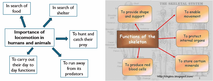

Without support, animals & humans would not be able to
maintain their body shape → their body will collapse
under the weight of their own tissues.
Support are provided by some form of skeleton :
Support are provided by some form of skeleton :
- Hydrostatic skeleton
- Exoskeleton, and
- Endoskeleton
Hydrostatic Skeleton (cecair)

- Consists of internal fluids (held under pressure in compartments surrounded by muscles).
- Since the liquid cannot escape, it forms a skeleton which cannot be compressed.
- This makes a soft-walled structure like an earthworm’s rigid so that muscles can act against it.
Exoskeleton (rangka luaran)

- Enclosed bodies of arthropods (insects, crustacean).
- Exoskeletons/ cuticle covers the body surface (cuticle - wax to prevent water loss)
- Support important internal organs & protect internal structure.
- Thin and flexible at
joints
- Enable organisms to move from place to place.
- Are non-living structures & incapable of growth → ecdysis (shed their exoskeleton)
Endoskeleton (rangka dalaman)

- Found in all vertebrates.
- Consists of rigid framework (made of bones and cartilage to which muscles are attached).
- Functions :
b). Supporting soft body tissue
c). Protecting internal organ from injury
- Certain parts store minerals, especially calcium and phosphorus, and produce blood cells.
Problems faced in support and locomotion
Problems that could be faced by humans and animals in
support and locomotion :
Aspect need to be considered when describing the locomotion of an animal :
- gravitational force, friction & resistance when moving around
Aspect need to be considered when describing the locomotion of an animal :
- Stability
– when it moves, it is temporarily unstable, but its stability will be restored
when it stops.
- Support – must have enough support from its body skeleton
How to overcome the problem?
- RESISTANCE & FRICTION – by streamlining their bodies.
- GRAVITATIONAL FORCE – most animals have their own supporting structures (fins – fishes, wings – birds)
- The skeletal system together with its muscles are designed specially to overcome the problems associated with support & locomotion of humans & animals.
- To initiate locomotion, the force required is generated by
contraction of muscle, whereas the movement is transmitted by changing of
skeleton position

Human Skeleton
- Consists mainly of bones.
- A few parts (nose, ears, soft disc between the vertebrae) made up of cartilage.
- 2 mains parts :
- Axial skeleton
- Appendicular skeleton
Axial Skeleton
Consists of :
- Skull
- Vertebral column
- Ribs
- Sternum (breastbone)
- Sacrum
- Coccyx
Appendicular Skeleton
Consists of :
- Pectoral girdle
- Pelvic girdle
- Upper limbs
- Lower limbs
Skull

- 22 bones
- Cranial bones – enclose & protect the brain
- Facial bones – protect &provide support for the entrance to the digestive and respiratory system
- Suture – held securely all skull bones.
- Jaw – the only freely
movable bone of the skull.
Vertebral Column
- Also known as spine/ backbone.
- Composed series of backbones called vertebrae (singular, vertebra)
- Functions :
- Encloses & protects the spinal cord
- Support the head
- Point of attachment
for the ribs, pelvic girdle & muscles of the back
- 7 cervical - movable
- 12 thoracic - movable
- 5 lumbar - movable
- 5 sacral/ sacrum – not movable
- 4
caudal/ coccyx – not movable
Ribs & Sternum (Thoracic Cage)

- Consists of ribs & sternum.
- Functions - Enclose
& protect organs in thoracic cavity & upper abdominal cavity
- 12 pairs of ribs make up the sides of the thoracic cavity (articulate with 12 vertebrae of the thoracic region)
- Sternum
– flat, narrow bone, located in the centre of the anterior thoracic wall.
The vertebrae (typical)
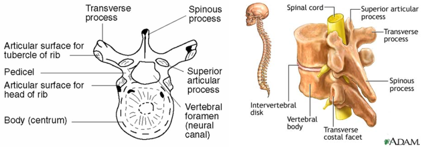
Cervical vertebrae
Thoracic vertebrae
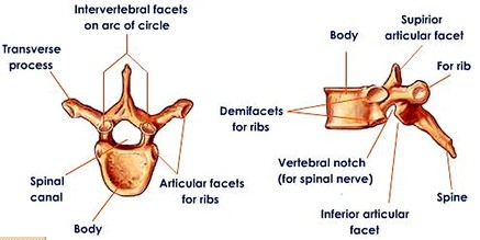
Spinous and transverse processes – serve as points of attachment for muscles & ligaments
Lumbar Vertebrae

Largest & strongest vertebrae. Their processes are short & thick. They also have large centrums which bear the weight of the lower back.
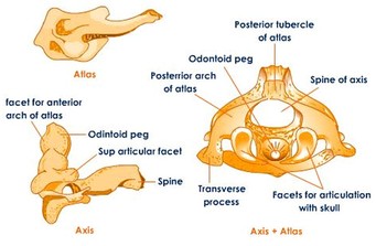
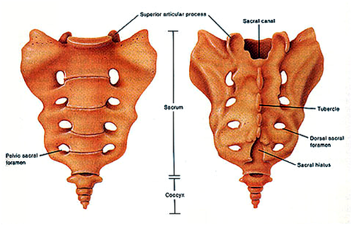
Sacrum & Coccyx
Appendicular Skeleton
Pectoral Girdle

Pelvic Girdle

Upper limbs

- 30 bones in each upper limbs.
- Humerus – longest & largest bone of the upper limb. It articulates with the scapula at the shoulder and with the radius (lateral bone) & ulna (medial bone) at the elbow.
- Carpus/Wrist - contains 8 short bones which are called the carpal bones. Each bone has a unique shape and name.
- Metacarpus/Palm - is supported by 5 long bones that are called metacarpals. Each bone is associated with a number starting with the thumb on the lateral side.
- Phalanges = bones of the
fingers. Each hand has 14 phalanges
Lower limbs

- Femur - longest, heaviest and strongest bone in the body. Head of the femur forms a ball-and-socket-joint with the hip bone. Other end of femur forms a hinge-joint with the tibia.
- Patella (kneecap) - Small triangular bone which protect the knee joint (hinge-joint).
- Tibia – bears the weight of the body.
- Fibula – smaller than tibia.
- Tarsus/ ankle – contains 7 bones called tarsals.
- Metatarsus – contains 5 bones of metatarsals.
- Phalanges
– similar to hand (number & arrangement)

LOCOMOTION IN ANIMALS
Earthworm

- Body wall has both longitudinal & circular muscles (act antagonistically to cause movement)
- Circular muscle contract and longitudinal muscle relax - causing segments to extend.
- Circular muscle relax and longitudinal muscle contract - causing segments to shorten.
- Chaetae secure the shorten segments to the ground (grip to the substratum).
Grasshopper


- The antagonistic muscle are flexor and extensor muscles.
- When the flexor muscle in the upper part of the leg is contracts, the lower leg is pulled towards the body.
- When the extensor muscle contracts (the flexor muscle relaxes), the hind legs jerks backwards propelling the grasshopper forward and upward into the air.
Fish


- A fish has an endoskeleton.
- its vertebral column is flexible and can be moved from side to side by the contraction and relaxation of antagonistic muscles called myotomes.
- When the muscles on one side of the fish's body contract, those on the other side relax.
- Alternating waves of contraction and relaxation pass down the myotomes on either side of the body from the head to the tail.
- These cause the different parts of the body to sweep from side to side, pushing the water backwards and sideways, and the body forwards.

To overcome instability during swimming :
- dorsal & ventral fins (median fins) - help fish to stay on course without yawing & rolling.
- paired pectoral & pelvic fins - to overcome pitching
- caudal/ tail fin - provide thrust and controls the direction.
Amphibian

- The hind legs are long and folded into a 'Z' shape when the frogs are at rest.
- Extensor muscle contract causing the hind legs straighten suddenly.
- The force produced is used to push the animal upwards and forwards.
- The frogs lands on its short forelimbs, which function to absorb shock of impact.
Bird


- Birds have aerofoil wings which generate the lift for flying through air.
- A pair of antagonistic muscles (pectoralis major and pectoralis minor) extend from the sternum to the humerus .
- Pectoralis minor muscles contract - wings are pulled up (upstroke)
- Pectoralis major muscles contract - wings are pulled down (downstroke)
Consequences of impaired musculoskeletal system on support and locomotion


